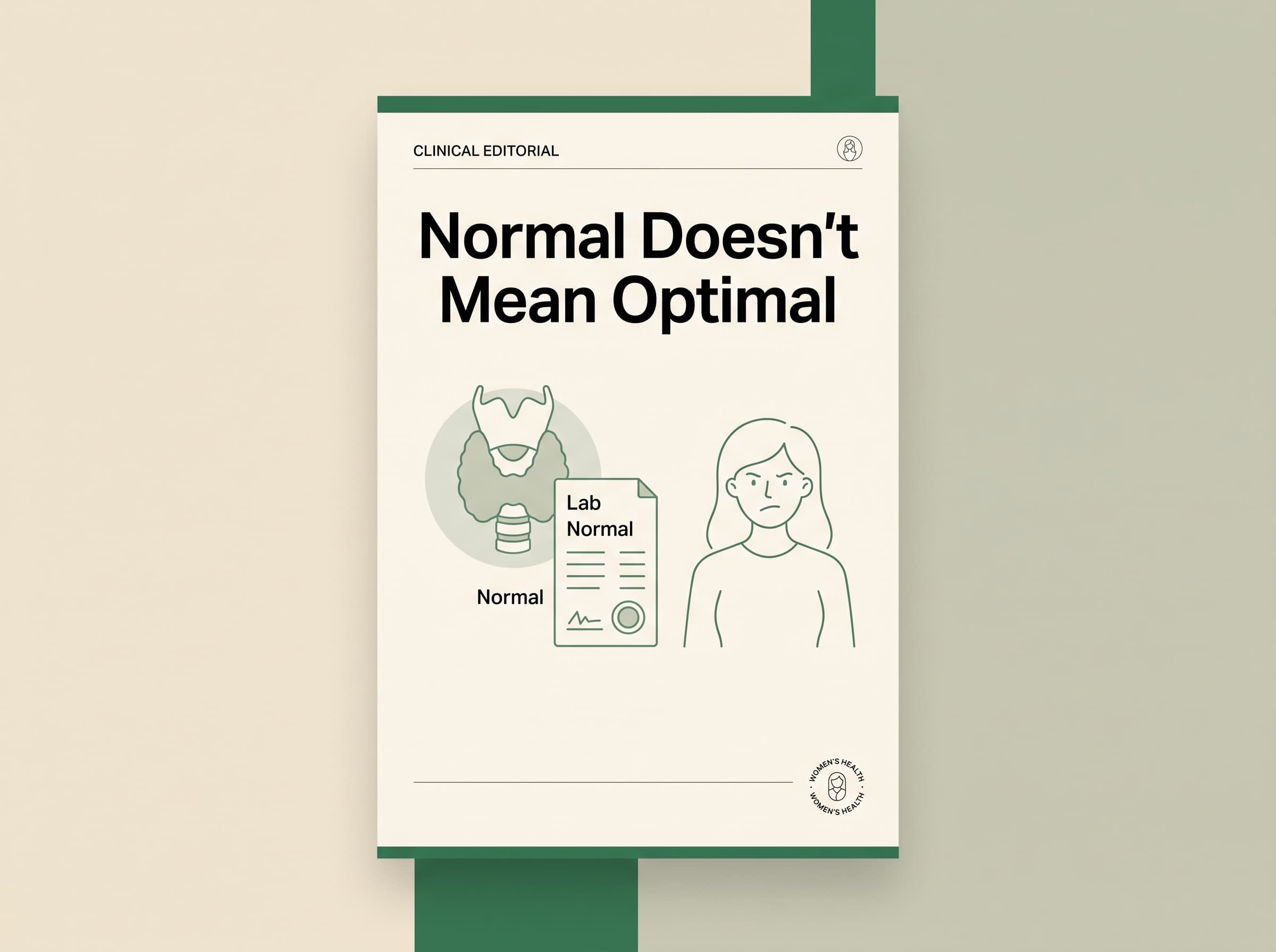
A normal TSH does not mean your thyroid is functioning optimally. It means your pituitary is working hard enough. Here is what the standard panel misses and what to ask for instead.
Why Your Thyroid Test Came Back Normal but You Still Feel Terrible
You went to your doctor with fatigue, weight gain despite no dietary change, hair thinning, constipation, cold hands and feet, brain fog, and depression that arrived without a life trigger. Your doctor ordered thyroid testing. The results came back normal.
And yet nothing has changed. The symptoms are still there.
You are not imagining them. You are also not getting the information you need from the tests that are being run.
Here is what is actually happening.
What TSH Measures (And What It Doesn't)
TSH stands for thyroid-stimulating hormone. It is produced by the pituitary gland, not the thyroid gland itself. The pituitary monitors thyroid hormone levels in the blood and adjusts TSH production to compensate for any deficiency.
When thyroid hormone levels fall, the pituitary produces more TSH to stimulate the thyroid to produce more. When thyroid hormone levels are adequate, TSH remains low because no additional stimulation is needed.
A normal TSH therefore tells you one thing: the pituitary is not in crisis mode. It is not compensating excessively. The system is, from the pituitary's perspective, maintaining adequate thyroid hormone levels in the blood.
What TSH does not tell you:
- How much active thyroid hormone is reaching your cells
- Whether the conversion from inactive T4 to active T3 is happening efficiently
- Whether Reverse T3 is blocking T3 from binding to thyroid receptors
- Whether your thyroid is under autoimmune attack
The Conversion Problem Nobody Talks About
Your thyroid primarily produces T4, an inactive form of thyroid hormone. T4 must be converted to T3, the active form that actually enters cells and sets metabolic rate, before it can do anything useful.
This conversion happens primarily in the liver, the gut (approximately 20% of T3 conversion occurs via gut bacterial enzymes), and peripheral tissues.
Multiple factors impair this conversion significantly:
- Low selenium: selenium is the critical cofactor for the deiodinase enzymes that convert T4 to T3. The majority of women are low in selenium.
- Chronic stress and elevated cortisol: cortisol directly suppresses T4 to T3 conversion and increases the production of Reverse T3.
- Gut dysbiosis: because a meaningful percentage of conversion happens in the gut, a dysbiotic gut produces less active T3 from the same T4 input.
- Caloric restriction: severe caloric deficit triggers the body to conserve energy by reducing T3 production, slowing metabolism.
- Systemic inflammation: inflammatory cytokines including IL-6 and TNF-alpha suppress T4 to T3 conversion at the enzyme level.
A woman can have a thyroid producing adequate T4, a normal TSH indicating the pituitary is satisfied, and still arrive at functionally insufficient active T3 reaching her cells because the conversion pathway is impaired. Her standard test will come back normal. Her symptoms will continue unchanged.
What Reverse T3 Is and Why It Matters
Reverse T3 is an inactive mirror-image form of T3. It is produced when T4 is converted in the wrong direction. Reverse T3 blocks T3 receptors on cells, preventing active T3 from binding and doing its job.
Reverse T3 production increases significantly under chronic stress, during illness, with severe caloric restriction, and with high-dose inflammatory states. It is essentially the body's emergency brake on metabolism.
A woman with high Reverse T3 can have normal TSH, normal Free T4, and even normal Free T3 on a standard panel, but still have impaired thyroid function at the cellular level because Reverse T3 is occupying the receptors that active T3 should be binding to.
Reverse T3 is not included in standard thyroid panels. It must be specifically requested.
Hashimotos: The Autoimmune Component
Hashimotos thyroiditis is the most common thyroid condition and the most common cause of hypothyroidism in developed countries. It is an autoimmune condition in which the immune system produces antibodies that attack thyroid peroxidase (TPO), the enzyme responsible for producing thyroid hormones.
The TSH can be completely normal for years, sometimes decades, while the immune system is actively attacking thyroid tissue. By the time TSH rises above the normal range, significant thyroid damage has often already occurred.
The diagnostic marker for Hashimotos is a positive TPO antibody test. This test is also not part of most standard thyroid panels. It must be specifically requested.
Women with Hashimotos also need to know that the autoimmune component of the condition responds to interventions that standard thyroid medication does not address. Gluten elimination (because molecular mimicry between gliadin and thyroid tissue drives antibody production in genetically susceptible individuals), selenium supplementation at 200mcg daily (the single most evidence-backed nutritional intervention for reducing TPO antibodies), gut healing (because intestinal permeability is a prerequisite for autoimmune activation), and vitamin D optimization all have meaningful evidence for reducing antibody levels and autoimmune activity.
The Complete Thyroid Panel Worth Requesting
The next time you discuss thyroid health with your doctor, ask specifically for:
Free T3: the active thyroid hormone that actually enters cells and sets metabolic rate. Not total T3, which includes bound and inactive forms. Free T3 specifically.
Free T4: the inactive precursor, to assess whether the thyroid is producing adequately and to understand the T4 to Free T3 ratio.
TSH: still useful as a reference point even if it is an incomplete picture on its own.
TPO antibodies: to identify the autoimmune component of thyroid dysfunction that may exist for years before TSH is affected.
Reverse T3: to identify whether the conversion pathway is producing excess inactive blocking hormone.
Ferritin: because ferritin below 70 ng/mL significantly impairs thyroid hormone production and energy even when thyroid markers appear normal on a panel.
Vitamin D: because thyroid receptor function is directly supported by adequate vitamin D levels, and deficiency is essentially universal in women above the 35th parallel.
What to Do With This Information
If your standard panel has come back normal but your symptoms persist, the conversation to have with your doctor is not "why don't you believe me?" It is a specific, named request for additional tests: Free T3, Reverse T3, TPO antibodies, ferritin, and vitamin D.
If your doctor is unwilling to order these tests, you have the right to request a referral to an endocrinologist. You also have the right to use functional medicine providers who routinely order comprehensive panels.
Your symptoms are data. The absence of an abnormal TSH does not make them less real. It makes the investigation more specific.
This article is for educational purposes only. Please work with a qualified healthcare provider for thyroid testing and treatment decisions specific to your health history.
More from Her Healthy Empire
The 10-Minute Habit After Meals That May Help Your Blood Sugar and Energy
A short walk after eating may do more than help you feel less sluggish. Research suggests post-meal movement can support the body’s glucose response, while adding an easy source of daily physical activity.
Why You’re Always Tired After 40, Even When You’re Getting Enough Sleep
Feeling exhausted even after a full night of sleep? Discover the overlooked reasons women over 40 may struggle with persistent fatigue, including sleep quality, perimenopause, iron, thyroid health, nutrition, stress, and more.
Period Changes in Perimenopause: What Is Normal?
Perimenopause can change cycle timing, flow, and duration. Some changes are expected, while unusual bleeding still deserves medical evaluation.
The next her healthy empire essay in your inbox.
One careful letter, every Sunday. Free.